Diagram Of The Muscles In The Forearm : 10 Best Images of Posterior Muscle Man Worksheet - Label ... - Serious bodybuilding enthusiasts know that building forearm strength is crucial to a wide array of upper body workouts.
Diagram Of The Muscles In The Forearm : 10 Best Images of Posterior Muscle Man Worksheet - Label ... - Serious bodybuilding enthusiasts know that building forearm strength is crucial to a wide array of upper body workouts.. Superficial muscles of the posterior forearm: Diagram the movements of the humerus muscles that act on the forearm. The superficial extensors of the forearm are the brachioradialis, extensor carpi radialis longus, anconeus, extensor carpi radialis brevis, extensor carpi ulnaris, extensor digitorum and extensor digiti minimi. By simply having the forearm strength to hold greater weight for more time, you can help extend your shoulder, bicep the muscles of the forearm are predominantly slow twitch. Remembering the action of each one can be quite difficult.
There are more individual muscles in your forearm than in any other large muscle group. Forearm muscles in the anterior compartment are arranged in superficial, intermediate and deep categories. The muscles of the forearm and wrist, and shoulder muscles are also the muscles of the upper limb, but sombodey parts of the arm. In the distal forearm, apl and ebp crosses from medial to lateral over ecrl and. Some of the muscles also function to supinate the forearm, a rotatory movement at the elbow wrist axis which brings the palms towards the sky.
Arm Muscle Diagrams from www.101diagrams.com The anconeus, located in the superficial region of the posterior forearm compartment, moves the ulna during pronation and extends the forearm at the elbow. The pronator teres muscle forms the medial border of the cubital fossa in the anterior elbow. Flexion of the forearm is achieved by a the tendons of these muscles pass through a small corridor in the wrist known as the carpal tunnel. Try labeling diagrams and worksheets as additional learning aids. Diagram the movements of the humerus muscles that act on the forearm. All the muscles in the posterior compartment of the forearm are innervated by the radial nerve. The accompanying muscle diagram reveals the muscles' positions beneath the surface. So, the muscles of the anterior compartment are generally innervated by the median nerve, with a few muscles being innervated by the ulnar nerve.
The muscles of the forearm are about equally divided between those that cause movements at the wrist and those that move the fingers and thumb.
4, attachment… the muscles of the back forearm. It is a functionally important muscle that contains two heads. The muscles of the upper arm are responsible for the flexion and extension of the forearm at the elbow joint. The forearm is the region of the upper limb between the elbow and the wrist. The antibrachial or forearm muscles may be divided into a volar and a dorsal group. Superficial muscles of the posterior forearm: The forearm is a mass of some 20 different muscles. Inflammation of this region caused by repetitive. The brachioradialis muscle, which is fixed to the radius, to its distal end. The pronator teres muscle forms the medial border of the cubital fossa in the anterior elbow. Try labeling diagrams and worksheets as additional learning aids. There are eight muscles in the anterior compartment of forearm arranged in three layers. Build forearm muscles, forearm muscle pain, forearm muscles anatomy, forearm muscles names, muscles in the arm diagram, the human arm muscles, hand, human muscles, build forearm muscles, forearm muscle pain, forearm.
It is a functionally important muscle that contains two heads. Pronator teres pronates the forearm, turning the hand posteriorly. Longus, brevis, longus, brevis (longus is lateral to brevis). This layer contains only one muscle, the flexor digitorum. Another handy relation to keep in the back of head is:
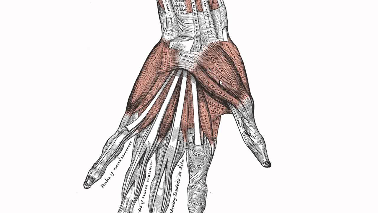
Muscles of the Hand - Anatomy Tutorial - YouTube from i.ytimg.com The forearm is a mass of some 20 different muscles. Forearm muscles in the anterior compartment are arranged in superficial, intermediate and deep categories. The term forearm is used in anatomy to distinguish it from the arm. This is the most medial of the superficial flexor muscles in the forearm. The flexor pollicis longus is situated on the radial side of the forearm, lying in the same plane as the preceding. Longus, brevis, longus, brevis (longus is lateral to brevis). Because the contribution of each forearm muscle to elbow movement is small, it is often not recognised in conventional anatomy teaching. Pronator teres pronates the forearm, turning the hand posteriorly.
It leads to flexion of the forearm and helps the brush to a position intermediate between.
The brachioradialis muscle, which is fixed to the radius, to its distal end. There are many muscles in the forearm. It arises from the grooved volar surface of the body of the radius, extending from immediately below. In the anterior compartment, they are split into three categories: Diagram of the muscles of the arm in action. The forearm is the region of the upper limb between the elbow and the wrist. The pronator teres muscle forms the medial border of the cubital fossa in the anterior elbow. The forearm is a mass of some 20 different muscles. Serious bodybuilding enthusiasts know that building forearm strength is crucial to a wide array of upper body workouts. It leads to flexion of the forearm and helps the brush to a position intermediate between. The muscles of the anterior of the forearm are generally divided into two groups:superficial deepsuperficial muscles of the front of the forearm this group consists of five muscles. It is a functionally important muscle that contains two heads. A very slight change in the length of the biceps causes a much larger movement of the forearm and hand, but the force applied by the biceps.
This layer contains only one muscle, the flexor digitorum. Inflammation of this region caused by repetitive. Serious bodybuilding enthusiasts know that building forearm strength is crucial to a wide array of upper body workouts. Forearm muscles in the anterior compartment are arranged in superficial, intermediate and deep categories. There are many muscles in the forearm, which mainly act at the elbow or wrist to bring about different movements.
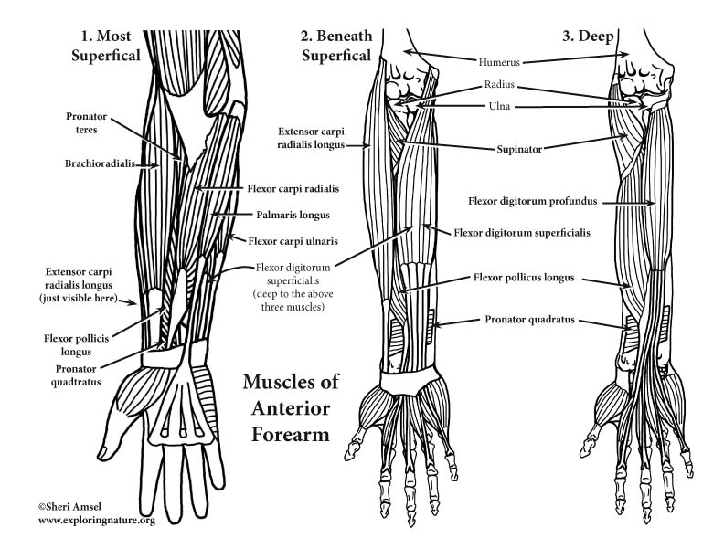
Muscles of the Arm and Forearm (Anterior) (Advanced) from www.exploringnature.org The forearm is the region of the upper limb between the elbow and the wrist. Longus, brevis, longus, brevis (longus is lateral to brevis). The anterior forearm muscles are divided into 3 muscular layers ; The antibrachial or forearm muscles may be divided into a volar and a dorsal group. Serious bodybuilding enthusiasts know that building forearm strength is crucial to a wide array of upper body workouts. A deep layer , intermediate layer and superficial layer. Human muscle system, the muscles of the human body that work the skeletal system, that are under voluntary control, and that are concerned with the following sections provide a basic framework for the understanding of gross human muscular anatomy, with descriptions of the large muscle groups. 2, ulna, 3, biceps muscle;
Build forearm muscles, forearm muscle pain, forearm muscles anatomy, forearm muscles names, muscles in the arm diagram, the human arm muscles, hand, human muscles, build forearm muscles, forearm muscle pain, forearm.
This is the most medial of the superficial flexor muscles in the forearm. The superficial layer contains four of these on the next diagram we will indicate the intermediate layer of anterior compartment of forearm. The forearm is the region of the upper limb between the elbow and the wrist. I've just switched over to a diagram to show you this muscle. The muscles of the upper arm are responsible for the flexion and extension of the forearm at the elbow joint. Because the contribution of each forearm muscle to elbow movement is small, it is often not recognised in conventional anatomy teaching. Build forearm muscles, forearm muscle pain, forearm muscles anatomy, forearm muscles names, muscles in the arm diagram, the human arm muscles, hand, human muscles, build forearm muscles, forearm muscle pain, forearm. The superficial extensors of the forearm are the brachioradialis, extensor carpi radialis longus, anconeus, extensor carpi radialis brevis, extensor carpi ulnaris, extensor digitorum and extensor digiti minimi. Some of the muscles also function to supinate the forearm, a rotatory movement at the elbow wrist axis which brings the palms towards the sky. Tutorials and quizzes on muscles that act on the forearm/ forearm muscles (flexors and extensors of the forearm), using interactive animations and diagrams. Inflammation of this region caused by repetitive. Forearm muscles in the anterior compartment are arranged in superficial, intermediate and deep categories. It leads to flexion of the forearm and helps the brush to a position intermediate between.


Posting Komentar
0 Komentar